Fluorescent Peptides for Research
Introduction
The advent platform of fluorescent peptides can enable advance research in cell system researches, no matter fluorescence or FRET energy transfer. Fluorescent peptides are the appropriate molecular probes, which are crucial to successfully interrogate complex cellular structure, and gain insight into molecule-molecule interactions, enzyme activities. This provides a better mechanism appearance of diseases formation and progression, and encourage clearer simulation of physiology and pathology in model cell systems.
Fluorescent labeling peptide is the novel chemical tools for molecular interactions in living organisms. Peptide-based fluorescent probes are advantageous over protein-based sensors, since fluorescent labeling peptide probes are synthetically accessible, easily modified and stable for selective biological applications with site-specific manners.
Fluorescent Peptide Labeling
The fluorophores in peptides can absorb light and re-emit as radiation, the emitted radiation has longer wavelength than the excitation light. Fluorescent labels (dyes) are bound to the amino-terminus of peptides in SPPS (solid-phase-peptide-synthesis), or site-specific linking to existing peptides. Moreover, thiol- or amine- groups in side-chains are also labeling without impact on biological functions. Biotinylated peptides can be labelled by fluorescent streptavidin- or avidin-conjugates.
All these fluorescent-labeling peptides are applied widely in biological researches as imaging studies and cellular enzyme assays. In the monitoring of protease activity, peptide substrates are labeled at the amino-terminus with the fluorophores, while the carboxy-terminus are attached with biotin moieties.
FRET Analysis
FRET applies the effective ability of fluorophores to label the molecules. The donor and acceptor are normally referred to as a FRET-pair. As there is no direct contact between fluorophore and quencher, the FRET takes place in the distal range between1-10 nm. This makes it suitable to measure process on the molecular scales.
Fluorescent Peptides in Medical Research
Fluorescent peptides are utilized to examine various disease states in medical researches, such as Alzheimer’s diseases, inflammation, autoimmune disease, cancer. This methodology can monitor the protease or cleavage in specific and defined manner, with base of different labeled peptide options and availability.
Molecular recognition
The molecular recognition of peptides or amino acids are critical factors in biochemical and medicinal process. Amino acids are the crucial as biosynthetic building blocks or signaling molecules, while peptides have functions as signaling molecules or hormones. Artificial peptide for molecular recognition has the following advantages:
- Peptides are comparatively small than proteins, and easily synthesized in the laboratory with standard protocols.
- The peptide sequence can be manipulated with precise AA functional groups for selective targeting.
- Peptides have enough size to contain the significant number of functional groups with precise location, all these amino acid sidechains can codify high-affinity and specific interactions with the target receptors.
Integration of specific peptide sequences with the organic fluorophores is the common method for the design of peptide-based fluorescent probes. Peptide-based environment-sensitive fluorescent probes are applied successfully in bio-molecule detection, these peptides can attach multiple fluorophores simultaneously as intrinsic module.
Biological Detection
Nucleoside Triphosphates Detection
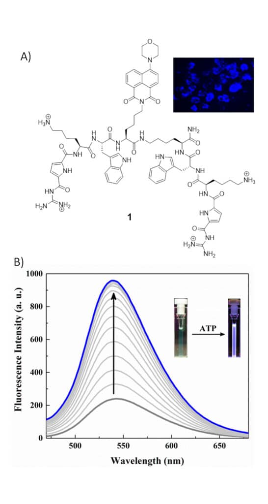
Nucleoside triphosphates (NTP) are involved in various biological processes, including cellular respiration, enzyme catalysis, energy transduction, and signaling. NTP is the most targeted anionic species, as its ubiquitous presence in the biological systems. There is a tweezer type peptide-based probe 1 from Schmuck and co-workers reports, this probe 1 can detect nucleoside triphoshpates in aqueous media.
Nucleic Acid Detection
Nucleic acids as the genetic information carrier exist in all living organisms, fluorescence monitoring of nucleic acids are critical in medical diagnosis, environmental monitoring, drug discovery, food safety.
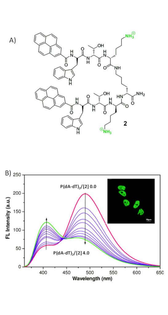
- The first fluorescent probe 2 is a pyrene-based peptide beacon, it has two Trp-Thr-Lys tripeptide arms attach to the central lysine spacer. The peptide beacon 2 exhibits typical pyrene-excimer emission at 490nm in folded form. Once bind to double-stranded DNA (dsDNA), it switches the fluorescence signal from excimer (490nm) to monomer emission (406nm). Therefore, monitor of relative fluorescence intensity at wavelength 406nm/490nm can detect the ratio-metric of nucleic acids.
- The cationic peptide beacon 3 can couple with different conjugates for ratio-metric detection of dsDNA. This peptide compound 3 has the naphthalene emission at 383 nm on 310 nm excitation, then emits at 535 nm upon 310 nm excitation once couple with dsDNA.
Both peptide beacons 2 and 3 based fluorescent probes provides a pronounced change in fluorescence signals for AT-rich polynucleotides than GC-rich polynucleotides. These fluori-metirc responses are also proportional to AT-base pair content in DNA. Moreover, both these two peptides can stain the nuclear DNA in research demonstrations.
Protein Detection
Specific Peptides and Insulin
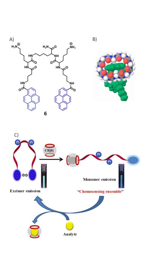
The combination of the pyrene-tagged amphiphilic peptide beacon 6 and a macro-cyclic host is applied for ratiometric fluorescent detection of amino acid derivatives, specific peptides, and proteins in aqueous media. The probe 6 emits pyrene excimer fluorescence at 505 nm with folded conformation in solution. Then CB additive can alter the fluorescence color change from pyrene excimer to monomer emissions. Therefore, probe 6-DB conjugates can be applied to monitor different substrates with affinity. Such as: screening methyl ester derivatives of hydrophobic amino acids (TrpOMe, PheOMe, LeuOMe), peptides with the hydrophobic amino acid residue at the N-terminus, human insulin.
14-3-3 Proteins
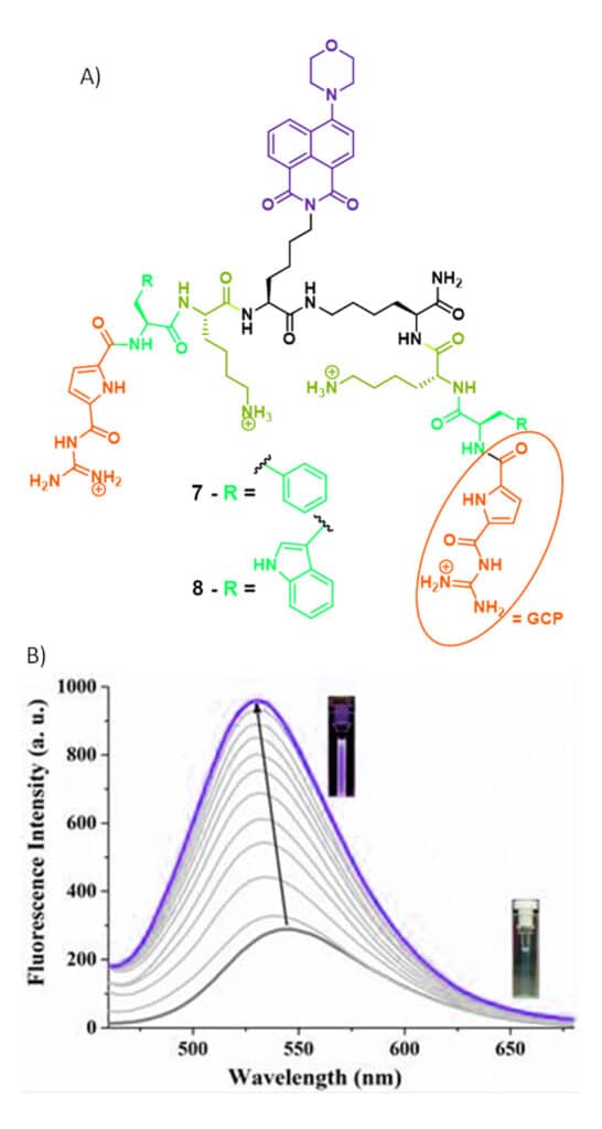
14-3-3 proteins has 7 isoforms of beta, epsilon, gamma, tau, theta, sigma, and zeta, this protein family has critical role in human physiology including: cell-cycle control, signal transduction, apoptosis, and neuro-transmission, metabolism, stress response, protein trafficking. There are two common fluorescent probes 7 and 8 for monitoring of 14-3-3 proteins. These peptide probes contain two symmetric GCP-Phe/Trp-Lys-Gly peptidic arms, and there is an artificial anion binding GCP moiety at both termini for 14-3-3 proteins recognition. In addition, there is an environment sensitive an aminonaphthimide fluorophore as fluorescence-based reporter.
β-Tryptase
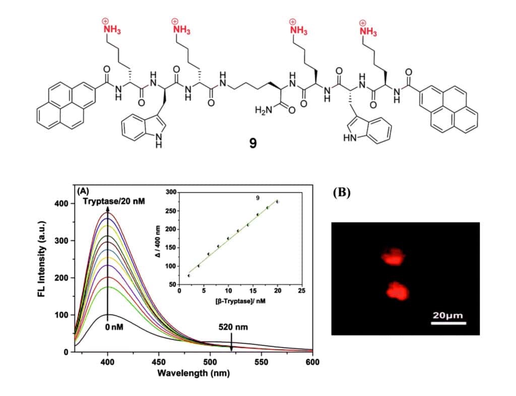
β-Tryptase is the predominant granule-derived serine protease, and secreted by human mast cells. It is involved in the pathogenesis of asthma, allergic and inflammatory disorders. The structure of this enzyme is tetramer, there are four identical subunits in two different orientations. There is a cationic fluorescent peptidic β-tryptase inhibitor, it has pyrene monomer emission at 400 nm and yrene excimer weaker emission at 520 nm. Once binding with β-tryptase, the monomer emission increases significantly, but excimer emission disappears. The enhancement of pyrene monomer fluorescence can indicate the binding between pyrenes and β-tryptase.
Heparin Detection
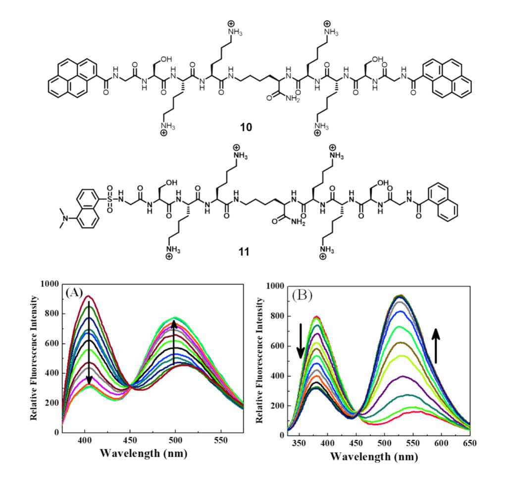
Heparin is a highly sulphated glycosaminoglycan, and has the highest negative charge density of bio-macromolecules. It is applied widely as both a prophylactic and a therapeutic agent, especially as an anticoagulant in surgery. The lysine-rich peptide beacons can target to a distinctive disaccharide unit of heparin for detection. The peptide beacon has two Lys-Lys-Ser-Gly tetrapeptide arms, and attach to the central lysine through C terminus. The heparin sensors 10 and 11 are designed by attaching the N-terminus with pyrenes and FRET pair respectively for ratiometric detection of heparin.
Lipopolysaccharide Detection
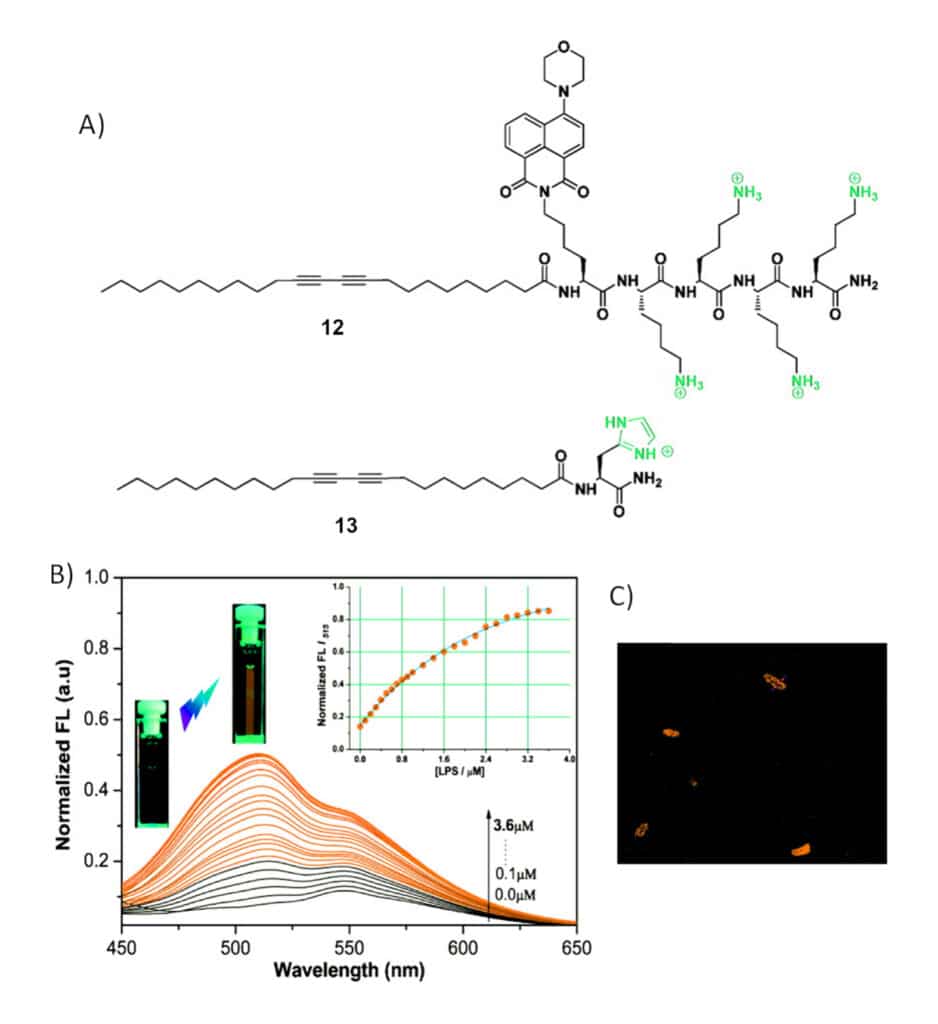
Endotoxic lipopolysaccharide (LPS) is a glyco-lipid, it is consisting of variable polysaccharide domain and lipid A, which is a conserved glucosamine-based phospholipid. LPS is an important component in the outer cell membrane of Gram-negative bacteria. There is a cationic peptide-based polydiacetylene (PDA) liposomes for fluorescent detection of LPS in mixed solvent.
The PDA liposomes alter colorless to red in formation of cross-linked, and the fluorescence of naphthimide is completely quenched. However, the addition of LSP will recover the fluorescence of the PDA liposomes to around 48% of the initial value. In addition, PDA liposomes are also applied for fluorescence staining of the membrane of E. coli bacteria.
Lysosomes Tracking
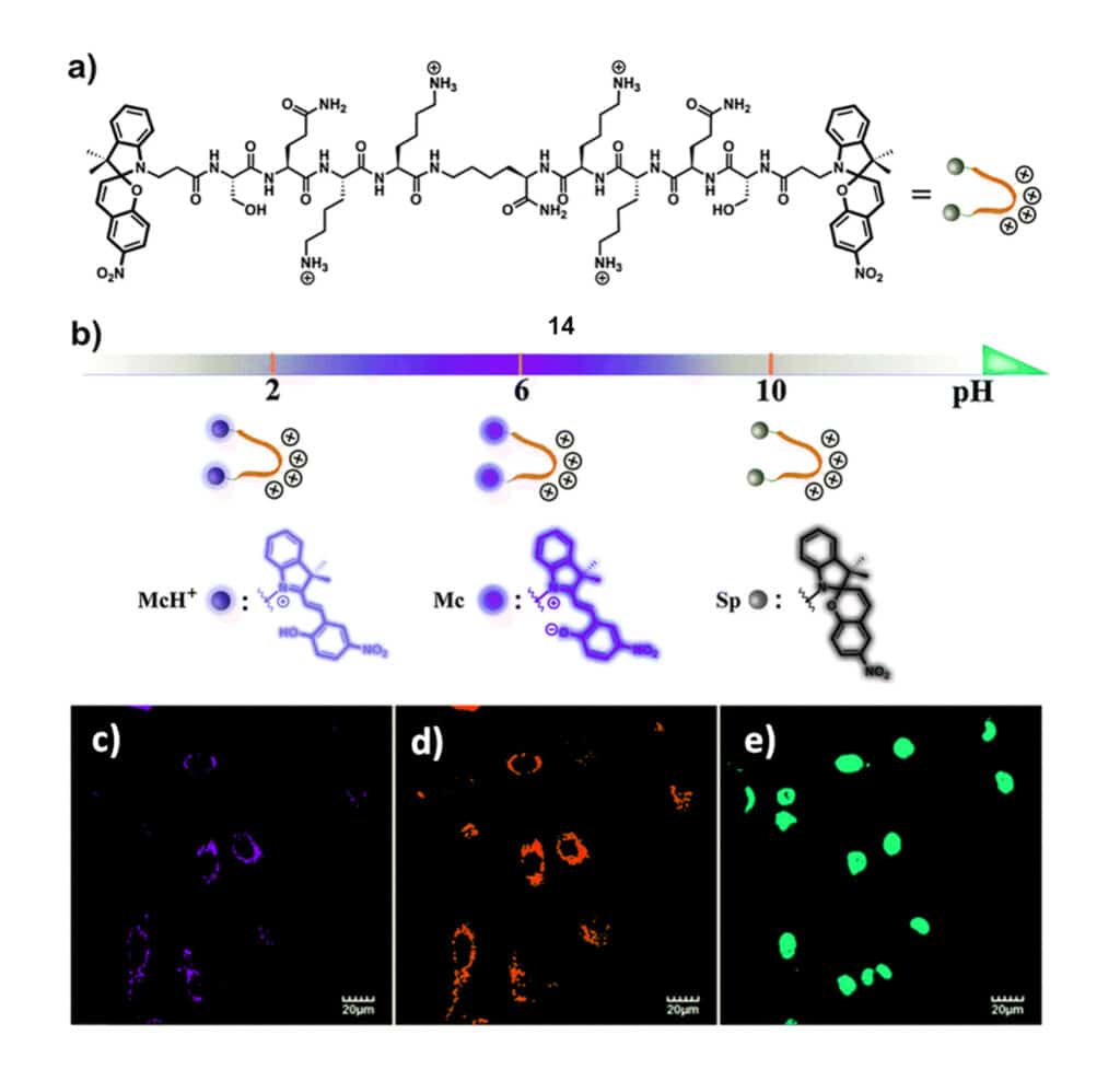
Lysosomes involves in multiple process like ntracellular transportation, metabolism, cell membrane recycling, and apoptosis. As deviation of lysosomal functions will result in diverse diseases, the close monitor and visualization are effective for the research of lysosome-related diseases. There is a bis-SP functionalized peptide 14 for pH monitoring in lysosomes and subsequent lysosomal imaging.
Conclusion
The development of fluorescent peptides is applied in different biological researches, Peptide-based probes based on FRET or fluorophores provide the basis of reliable design strategies of various applications, including analytic detection, molecular interaction of protein-peptide and protein-protein. The novel fluorophores and tailor-made receptors open the possibility of new peptide-based chemical tools for biological analysis and clinical diagnosis.
Contact Qyaobio with novel fluorescent peptides for your new biological researches.


